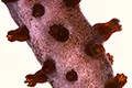
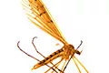
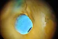
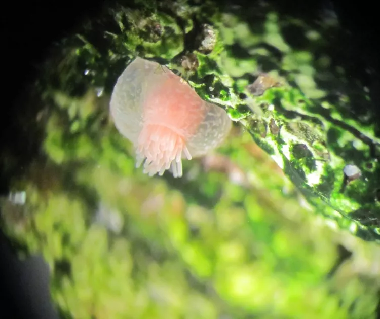
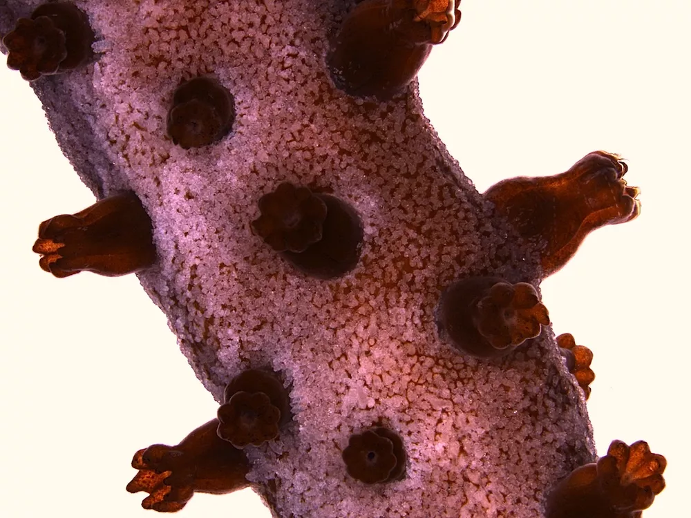
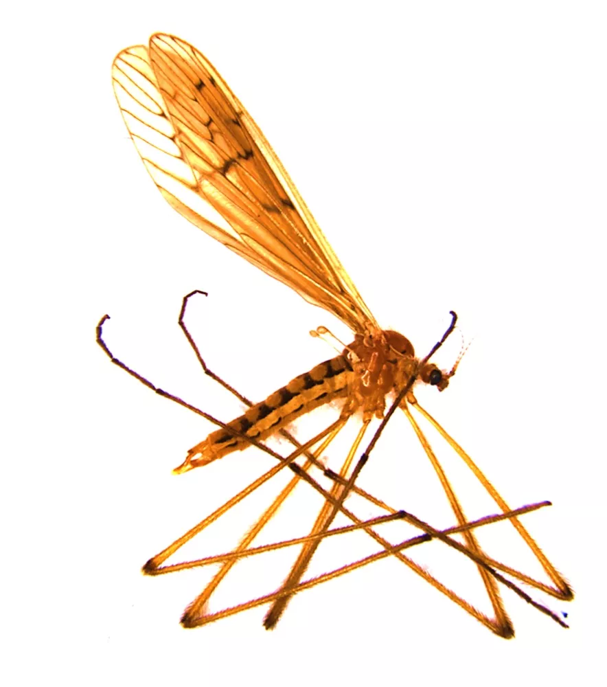
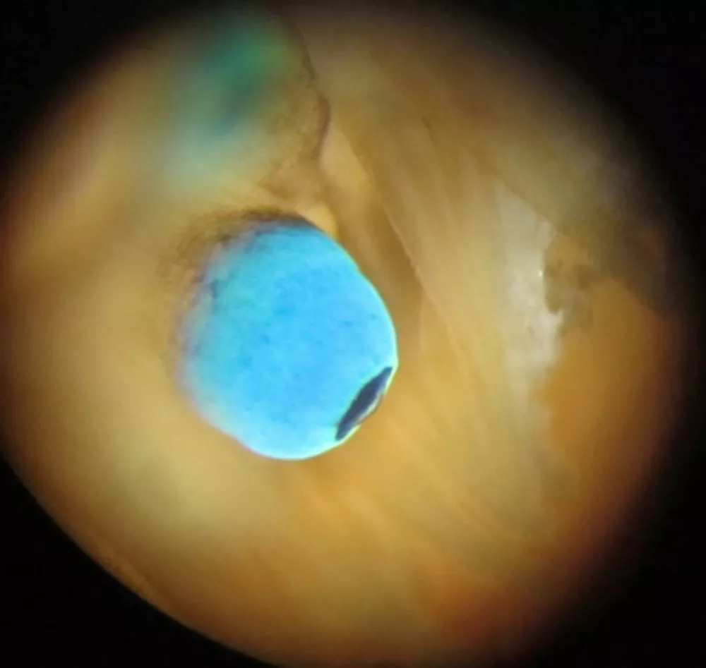
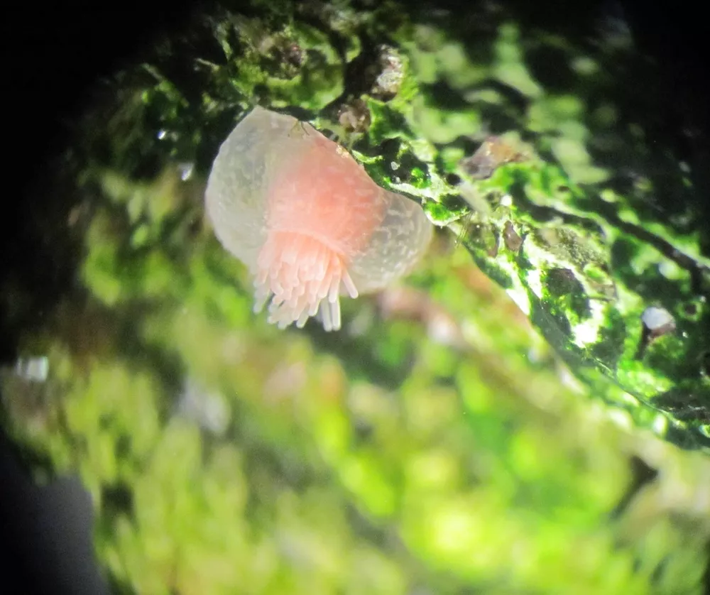
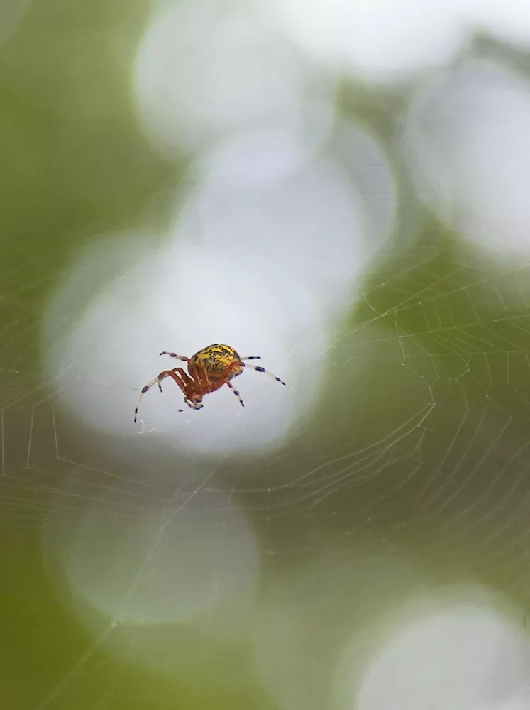
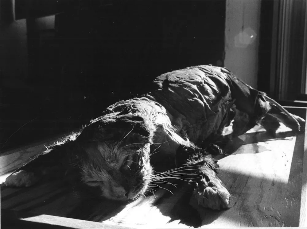
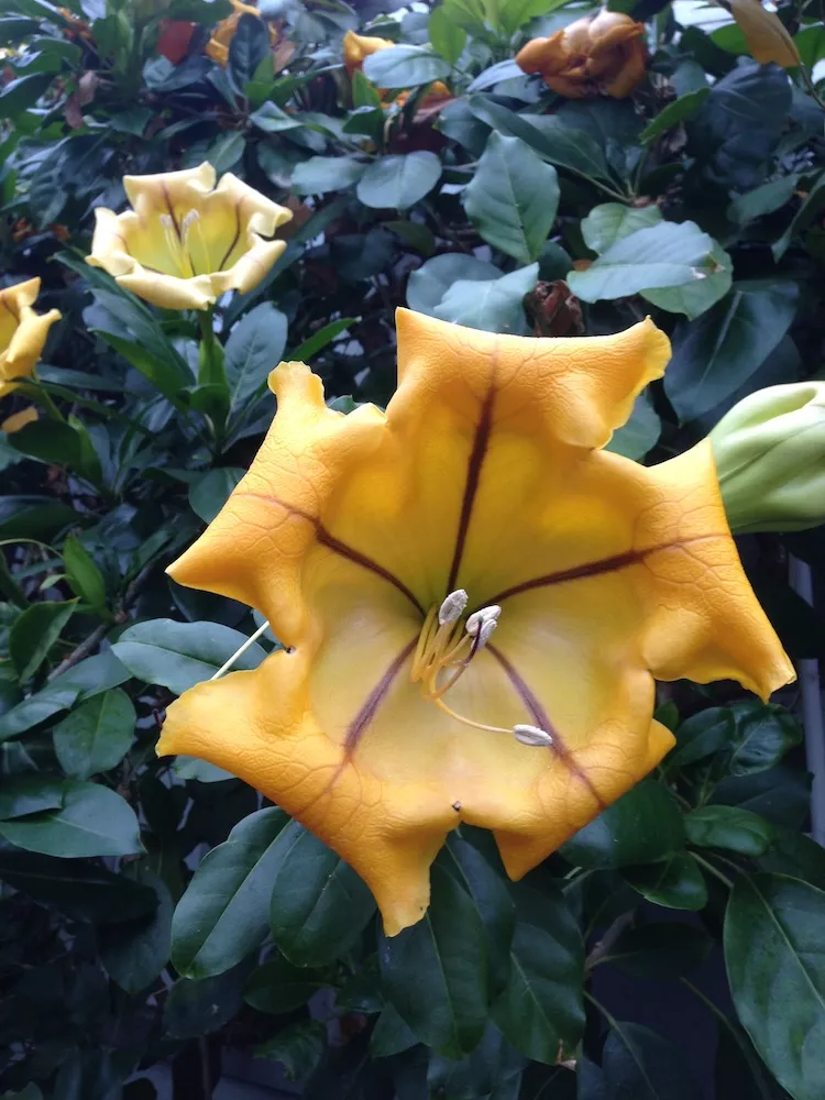
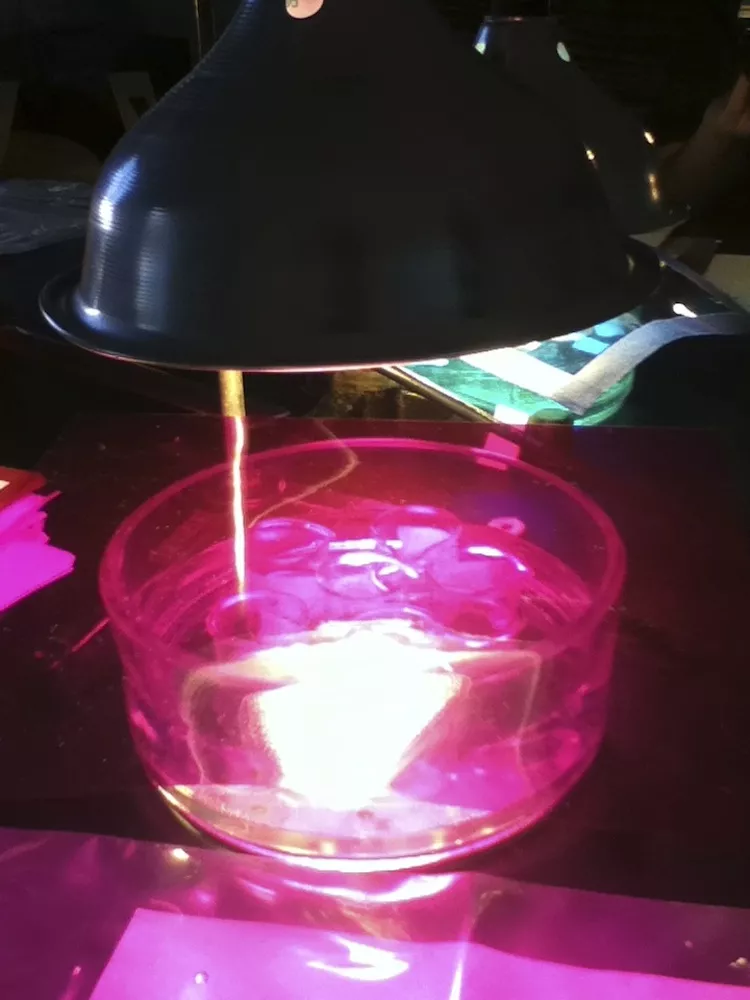
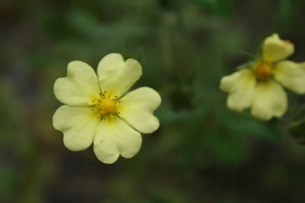
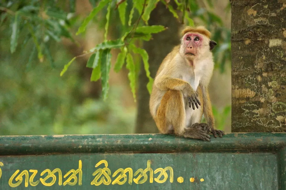
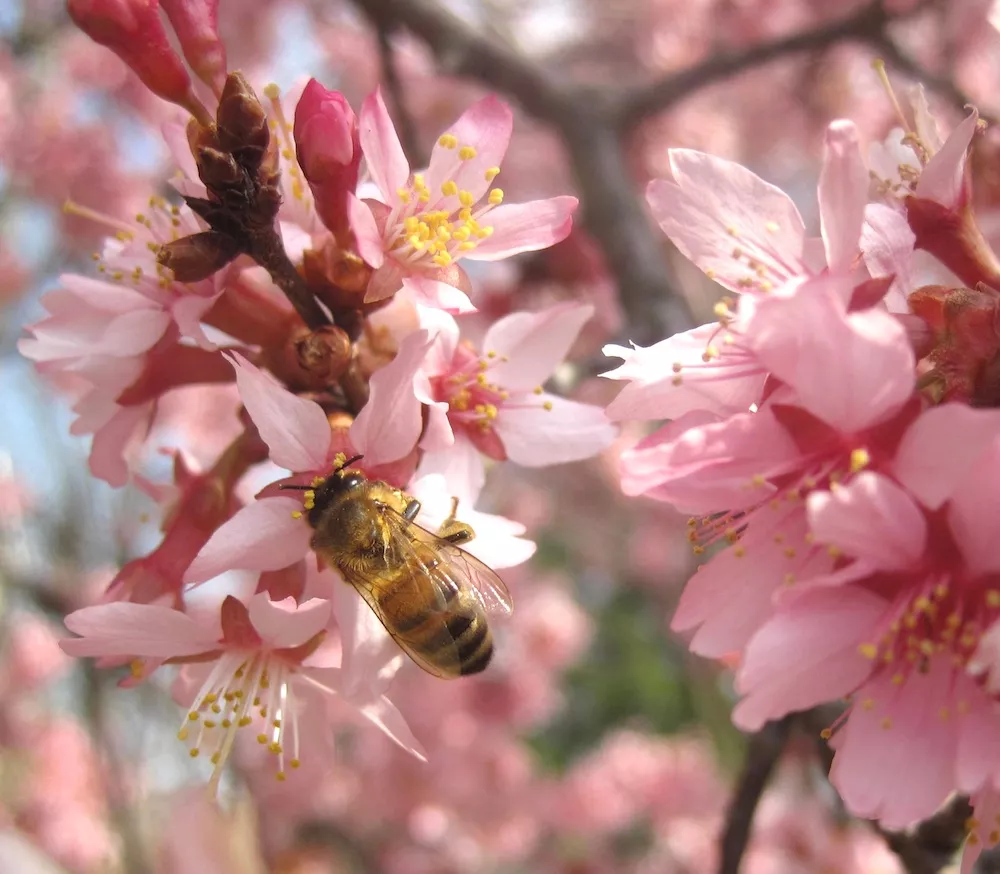
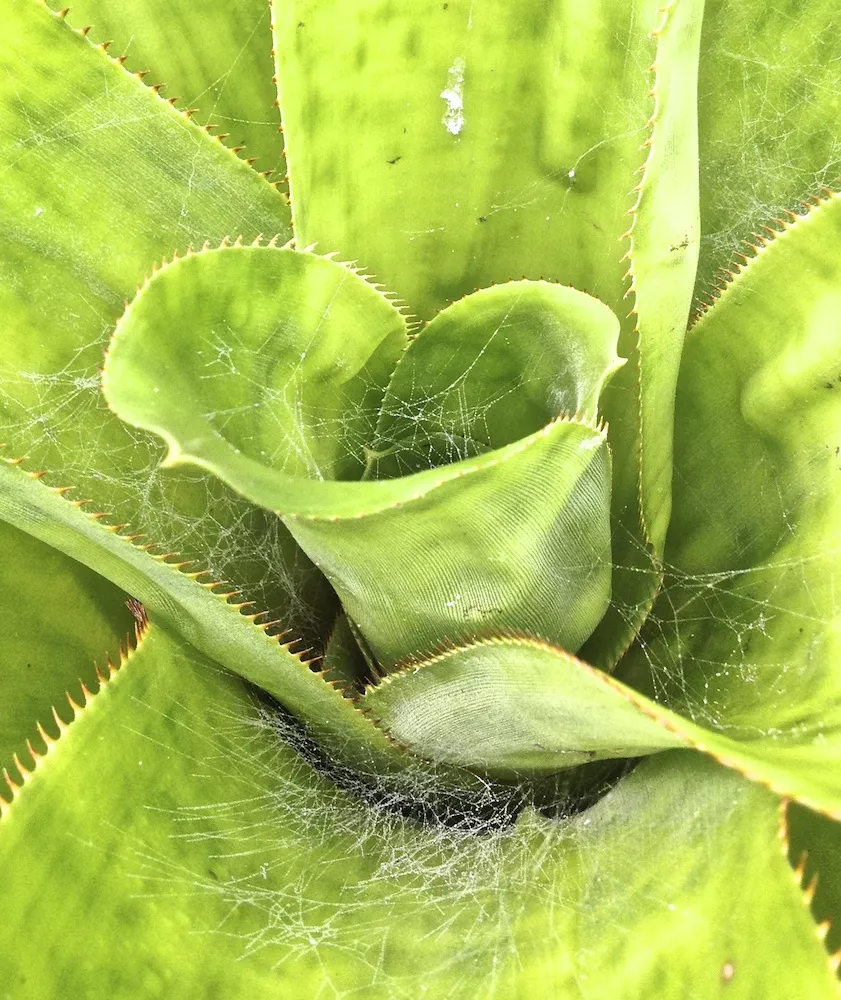
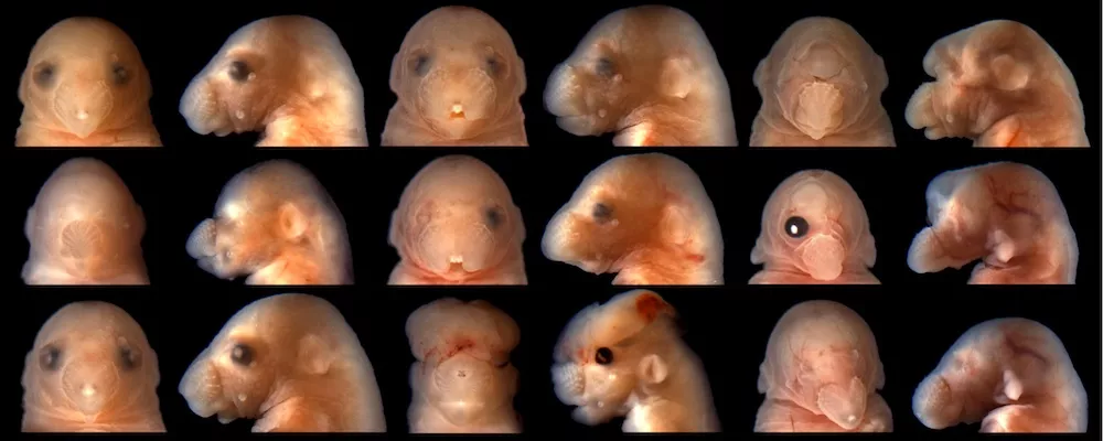
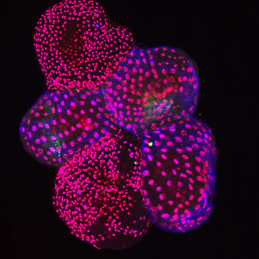
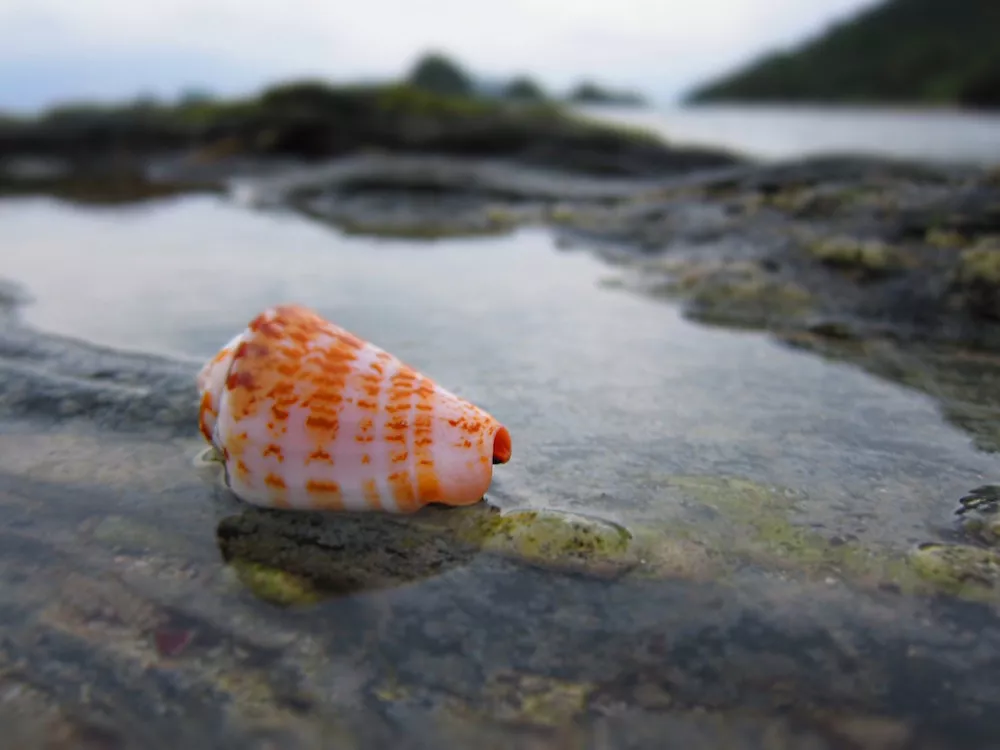
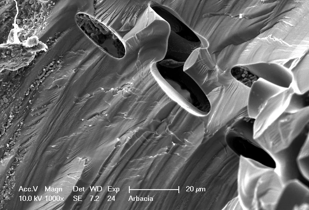
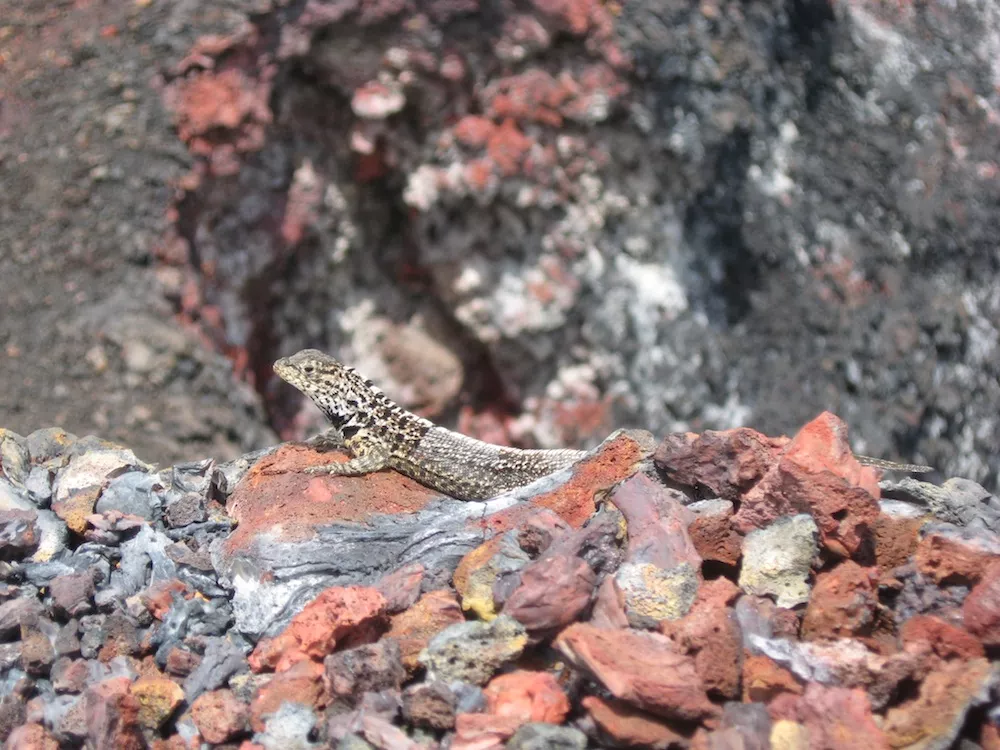
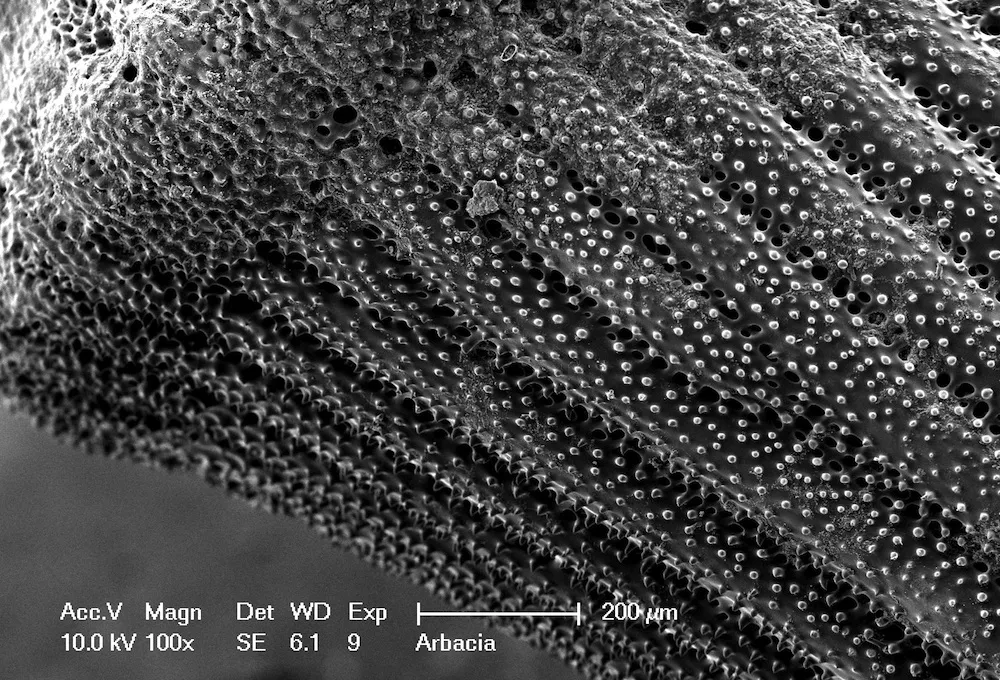
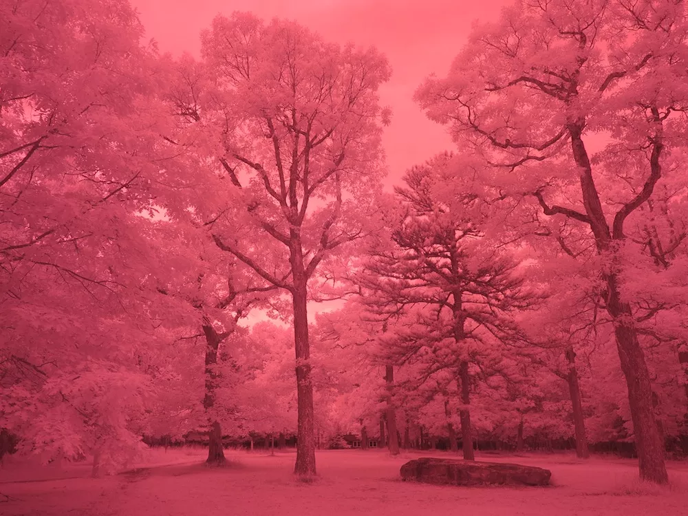
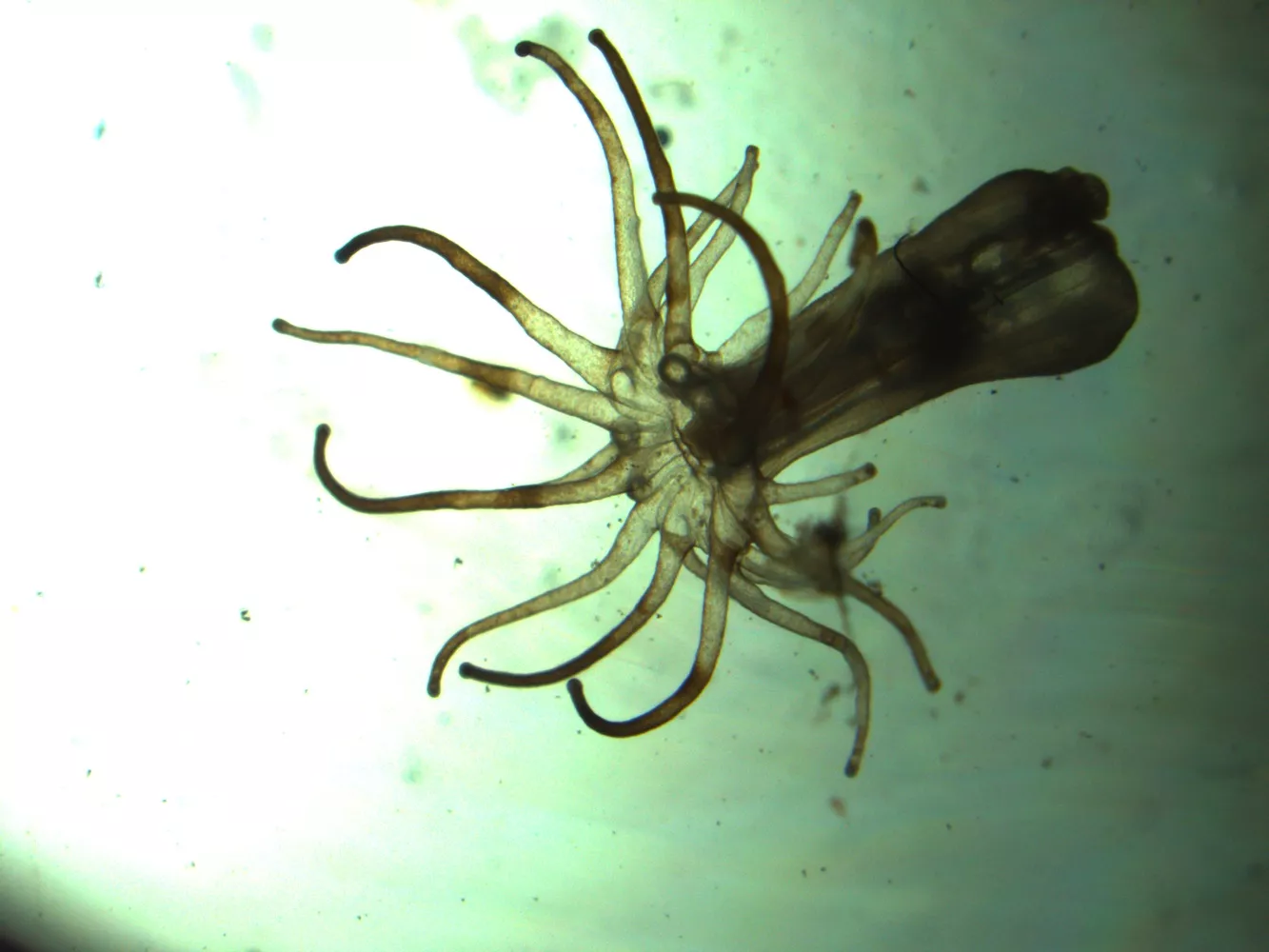
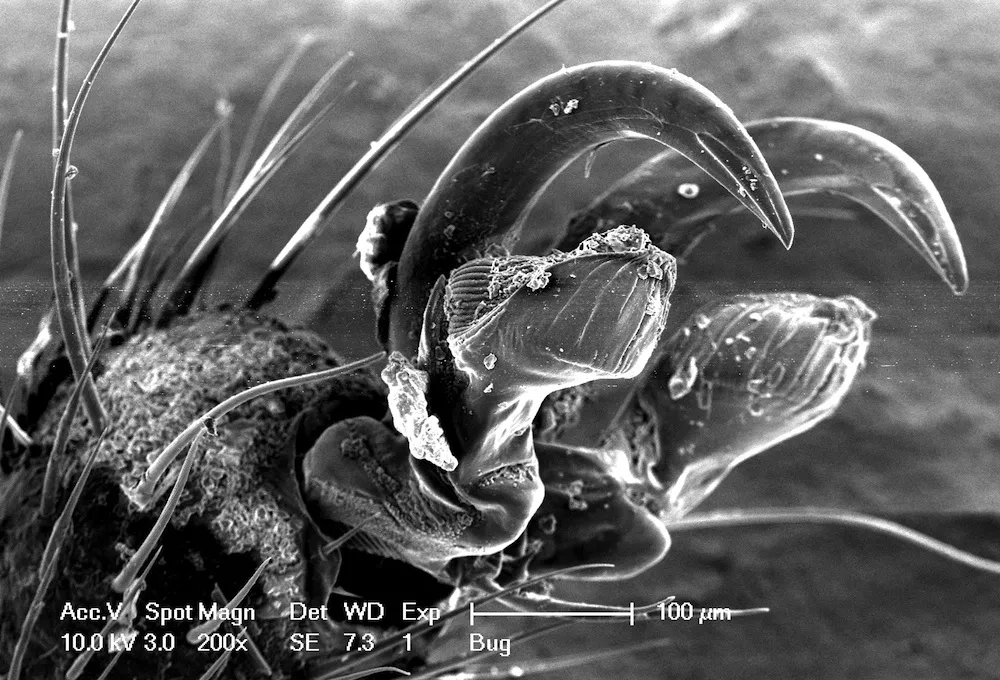
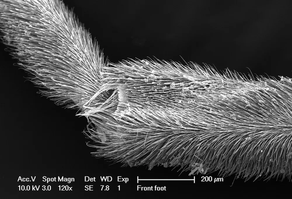
Slideshow
![An octocoral of the genus Leptigorgia](/sites/default/files/styles/image_slider/public/assets/images/news-events/fs_savage13_12.jpg.webp?itok=k3YbFC57)
First Place (TIE) - An octocoral of the genus Leptigorgia. Octocorals – commonly referred to as “sea whips” or “sea fans” for their delicate and elaborate branching structure – are an abundant form of colonial cnidarian. Two features are worth noting: first are the beautiful crystals that coat the surface of the central axial rod – these calcareous structures are called spicules and provide protection for the soft tissue of the coral within. Second is the deep brown pigmentation of the emerging coral polyps – the coloring comes not from pigment cells but from millions of zooxanthellae, a type of algae, living symbiotically in the coral. The image was captured using an extended depth of field microscope.
Photo
by
Erin Kast '15<br>religion major<br>Wausau, Wis., and<br>Raul Anchiraico '14<br>biology major<br>Sugar Land, Tex.
![Crane flies](/sites/default/files/styles/image_slider/public/assets/images/news-events/fs_savage13_08.jpg.webp?itok=GxZJuwJQ)
First Place (TIE) - Crane flies belong to the order of Diptera. Common members of Diptera include mosquitos and flies. This crane fly is found hanging out on the wall of my dorm. Its back-wing, as showed in the picture, has degenerated into a pair of lobes. If you ever see one of these crane flies, don’t panic and misled them as huge mosquitos. They don’t bite and mostly feed on nectars. The picture could also be used to train your ten-year-old nature-loving cousin, just ask him to figure out what is not right with this picture. (Hint, insects belong to the superclass Hexpoda.)
Photo
by
Zhengyang Wang '14<br>biology and philosophy major<br>Chengdu, China
![Scallops (family Pectinidae) have the most acute visual sensory organs of any bivalves.](/sites/default/files/styles/image_slider/public/assets/images/news-events/fs_savage13_21.jpg.webp?itok=WZVetqtm)
Second Place - Scallops (family Pectinidae) have the most acute visual sensory organs of any bivalves. An individual scallop will usually have an array of 40-60 eyes along its mantel cavity, with some species possessing over 100! These eyes are capable of detecting the approach of predators, such as sea stars, sharks, and other fishes. They may also be used by swimming scallops to pick out favorable substrates to come to rest upon. This scallop was viewed live under a light microscope at 16x magnification. Image taken using an 8x digital camera.
Photo
by
Patrick Ammerman '14<br>biology major<br>Cincinnati, Ohio
![Anthozoan](/sites/default/files/styles/image_slider/public/assets/images/news-events/fs_savage13_05.jpg.webp?itok=-_j68Gkl)
Third Place - This image depicts the Anthozoan, Aiptasiomorpha sp., in its retracted state after experiencing physical stimuli. It caves its tentacles into the gastrovascular cavity in order to minimize the amount of volume exposed to potential predators. The sea anemone was ~2 cm from the base to tip of the mouth (in its ‘extended’ state). The picture was taken through the lens of a dissecting scope.
Photo
by
Justin Sui '15<br>biology major<br>Yardley, Pa.
![The harmless marbled orb weaver](/sites/default/files/styles/image_slider/public/assets/images/news-events/fs_savage13_01.jpg.webp?itok=sUhI4xZM)
The harmless marbled orb weaver (Araneus marmoreus var. pyramidatus) in the Crum Woods on a fall afternoon.
Photo
by
Julia Carleton '15<br>Wyoming, Ohio
![This cat was pregnant and being looked at anatomically for my Comparative Vertebrate Anatomy course.](/sites/default/files/styles/image_slider/public/assets/images/news-events/fs_savage13_22.jpg.webp?itok=m7IH1dLv)
This was taken with 35 mm film. This cat was pregnant and being looked at anatomically for my Comparative Vertebrate Anatomy course. Thanks, Jason!
Photo
by
Alexis Dziedziech '13<br>psychology and biology major<br>Freeport, N.Y.
![A flowering shrub](/sites/default/files/styles/image_slider/public/assets/images/news-events/fs_savage13_23.jpg.webp?itok=RnN049wF)
A flowering shrub, seen at the historic Longwood Gardens
Photo
by
Michael McVerry '16<br>Vienna, Va.
![Six beakers of Ilex opaca (Holly) leaf disks in sodium bicarbonate solution under a purple light.](/sites/default/files/styles/image_slider/public/assets/images/news-events/fs_savage13_02.jpg.webp?itok=W9CRXpEG)
Six beakers of Ilex opaca (Holly) leaf disks in sodium bicarbonate solution under a purple light. Taken during a Bio 2 lab measuring net rate of photosynthesis under different wavelengths of light. The idea was that leaf disks infiltrated with solution (which makes them sink) would produce oxygen bubbles as photosynthesized and rise to the surface.
Photo
by
Azucena Lucatero '16<br>Ontario, Calif.
![cinquefoil flower](/sites/default/files/styles/image_slider/public/assets/images/news-events/fs_savage13_03.jpg.webp?itok=uKxWcRAz)
Hiking on a grey morning in the Laurentian Mountains in Quebec, I came across this cinquefoil flower (genus Potentilla, most likely Potentilla recta), its muted yellow a stark contrast to the wet green of its leaves. Its withering sister behind it only drew more attention to perfect symmetry of the living flower, itself a small testament to the vibrant geometry found in the natural world.
Photo
by
Maddy Booth '15<br>Brentwood, Tenn.
![toque macaque](/sites/default/files/styles/image_slider/public/assets/images/news-events/fs_savage13_04.jpg.webp?itok=nSfqRytn)
The endangered toque macaque (Macaca sinica) monkey, endemic to Sri Lanka. Photo taken at Sri Lanka’s Yala National Park.
Photo
by
Daniela Kucz '14<br>biology & gender & sexuality studies major<br>Singapore
![A honeybee pollinating cherry blossoms in the Arboretum Cherry Border](/sites/default/files/styles/image_slider/public/assets/images/news-events/fs_savage13_06.jpg.webp?itok=Ou2y8F0j)
A honeybee pollinating cherry blossoms in the Arboretum Cherry Border is one of prettiest examples of coevolution. A similar sight inspired Darwin, who wrote in The Origin of Species, “Thus I can understand how a flower and a bee might slowly become… adapted in the most perfect manner to each other.” This coevolution continues today, but since Darwin’s time human beings have changed the selection pressures on both bees and ornamental trees.
Photo
by
Phoebe Cook '15<br>Adamant, Vt.
![Icicles resemble a vector field](/sites/default/files/styles/image_slider/public/assets/images/news-events/fs_savage13_07.jpg.webp?itok=zDEnDEUs)
Icicles resemble a vector field indicating the presence of high speed winds in an alpine biome (Mt. Adams in the White Mountains, NH).
Photo
by
Nick Sohn '15<br>engineering major<br>Mill Valley, Calif.
![a spider web within a green Aechmea plant](/sites/default/files/styles/image_slider/public/assets/images/news-events/fs_savage13_09.jpg.webp?itok=poGR3qi1)
This is a close-up photograph of a spider web within a green Aechmea plant at Longwood Gardens. Not pictured is the industrious spider who took advantage of the rosette foliage of the Aechmea plant, a large bromeliad.
Photo
by
Cecilia Paasche '16<br>Bronx, N.Y.
![fetus](/sites/default/files/styles/image_slider/public/assets/images/news-events/fs_savage13_10.jpg.webp?itok=LKF3yupY)
This is a figure compiled from my summer research in a fetal toxicology lab at UNC. This image encompasses the various effects of gene-environment interactions resulting the combination of fetal alcohol exposure and genetic heterozygosity. These mice fetuses were imaged using a light microscope and vary in phenotype from median cleft lip to anophthalmia.
Photo
by
Henry Kietzman '14<br>neuroscience major<br>Mystic, Conn.
![propidium iodide-stained DNA](/sites/default/files/styles/image_slider/public/assets/images/news-events/fs_savage13_11.jpg.webp?itok=xNhXRaAL)
Tiny red dots of propidium iodide-stained DNA light up the nuclei of Ciona intestinalis embryos, both in the follicle cells of two chorion-coated embryos and the epidermal nuclei of three dechorionated embryos, all fixed in methanol. Blue tubulin marks mitotic spindles while slight green stain highlights fibroblast growth factor receptor in now-defunct heart progenitor cells developing just under the outer surface of the embryos. Image taken with a Leica SP5 confocal microscope and composite Z-stacks were created in ImageJ.
Photo
by
Emily MacDuffie '13<br>biology major<br>Cape Elizabeth, Maine
![abandoned cone snail shell](/sites/default/files/styles/image_slider/public/assets/images/news-events/fs_savage13_13.jpg.webp?itok=Hmok8KWQ)
An abandoned cone snail shell found on a beach in Okinawa, Japan. Cone snails can have quite a venomous sting, and are known to hospitalize many people in the area on an annual basis.
Photo
by
Patrick Monari '13<br>biology and psychology major<br>Rutledge, Penn.
![Side view of the fractured region of a bottom spine from the sea urchin](/sites/default/files/styles/image_slider/public/assets/images/news-events/fs_savage13_14.jpg.webp?itok=dIY3octX)
Side view of the fractured region of a bottom spine from the sea urchin, Arbacia punctulata, under a scanning electron microscope. The striated regions mark the inside walls of the spine, and the largely porous nature of these spines are indicated by the holes.
Photo
by
Andrew Han '14<br>biology and psychology major<br>Seoul, Korea
![A Galapagos lava lizard at the mouth of El Chico volcano on Isla Isabela in the Galapagos](/sites/default/files/styles/image_slider/public/assets/images/news-events/fs_savage13_15.jpg.webp?itok=91lLf6hP)
A Galapagos lava lizard at the mouth of El Chico volcano on Isla Isabela in the Galapagos. This photo captures a male doing “push-ups” to mark and defend his territory. Of the seven species of lava lizard endemic to the Galapagos, the Galapagos lava lizard is the only found on multiple islands (in the central and western archipelago), suggesting that these islands were more recently connected than their eastern counterparts.
Photo
by
Yvonne Socolar '13<br>biology and psychology major<br>Chapel Hill, N.C.
![Base of a sea urchin spine](/sites/default/files/styles/image_slider/public/assets/images/news-events/fs_savage13_16.jpg.webp?itok=MhUW61_c)
Base of a sea urchin spine taken from Arbacia punctulata with an scanning election microscope.
Photo
by
Kai Tucker '13<br>biology major<br>Los Angeles, Calif.
![Infrared photograph of the mixed forest](/sites/default/files/styles/image_slider/public/assets/images/news-events/fs_savage13_17.jpg.webp?itok=ZlDYlTUu)
Infrared photograph of the mixed forest at the Mountain Lake Biological Station located deep within the Appalachian Mountains in Giles County, Virginia. In the background is the Laing Dormitory where researchers live during the summer months. The white foliage is due to chlorophyll reflecting infrared light in what is know as the “Wood Effect” after physicist Robert Wood. The photograph was take with a Canon PowerShot G1X converted to utilize light in the infrared spectrum for nocturnal research.
Photo
by
Daniel Hess '13<br>religion and biology major<br>Greencastle, Penn.
![A specimen of aiptasia pallida](/sites/default/files/styles/image_slider/public/assets/images/news-events/fs_savage13_18.jpg.webp?itok=XDfn4BHd)
A specimen of aiptasia pallida with its algea symbiont bleached out of its cells.
Photo
by
Valentina Garcia '14<br>biology major<br>Oakland, Calif.
![tarsal claw at the tip of a Stink bug’s foot](/sites/default/files/styles/image_slider/public/assets/images/news-events/fs_savage13_19.jpg.webp?itok=WYOzi7oL)
The photo shows the small tarsal claw at the tip of a Stink bug’s foot (Family pentatomidae, Genus Halyomorpha.) The photo was taken with a scanning electron microscope with a magnification of 200x.
Photo
by
Geli Carabases '14<br>economics major<br>Mount Laurel, N.J.
![Photo of Water Strider’s leg covered in small hydrophobic hairs](/sites/default/files/styles/image_slider/public/assets/images/news-events/fs_savage13_20.jpg.webp?itok=XPWV29ku)
Photo of Water Strider’s leg covered in small hydrophobic hairs. When observing the hairs closely, we see they are covered in small grooves, giving them their hydrophobic nature. A “comb” like structure of the exoskeleton can be seen near the joint. This is used to comb clean the hairs and keep the water striders afloat. The picture was taken with a scanning electron microscope at 120x magnification.
Photo
by
Corinne Sommi '14<br>biology major<br>Darien, Conn.
1
of
23