B. 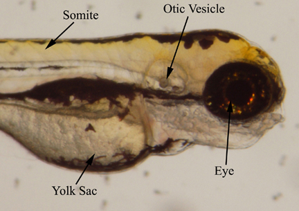
C. 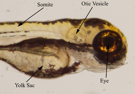
B.
C.
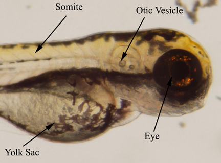
Figure 5. Anterior region of zebrafish embryos approximately
70 hours after treatment with retinoic acid. No apparent
abnormalities are seen in the embryos in the 10-11 M (B) and 10-10 M
(C) solutions when compared with the control (A). Eye, otic vessicle,
and somite development appear very similar. The heads of the embryos
treated with 10-11 M (B) and 10-10 M (C) solutions appear to be
slanted backwards.