B. 
C. 
D. 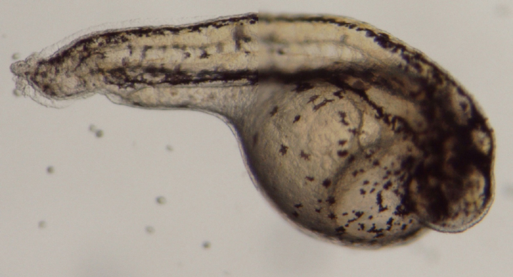
B.
C.
D.
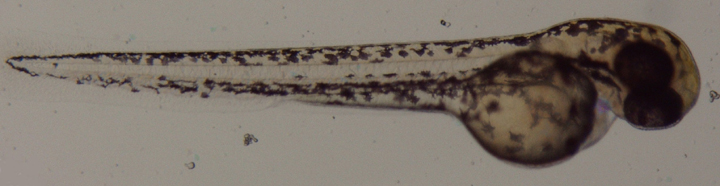
Figure 3. Zebrafish embryos approximately 46 hours after treatment with retinoic acid. Control embryos (A) and those in the 10-11 M (B) and 10-10 M (C) solutions continue normal development. The embryos in the 10-8 M (D) solution continue to develop, but at a slower rate. Melanocytes migrated posteriorly in the embryos, but not in the same regular pattern for the embryos in the 10-8 M, as for the control and those in the 10-11 M and 10-10 M solutions.