
B. 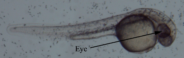
C. 
D. 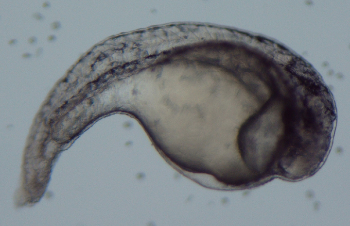
B.
C.
D.
Figure 1. Dechorionated zebrafish embryos approximately 24 hours after treatment with retinoic acid solutions. Compared to the development of the control embryos (A), development of the embryos in the 10-11 M (B) and 10-10 M (C) retinoic acid solutions seems to have proceeded normally. Embryos treated with retinoic acid at a concentration of 10-8 M (D), however, exhibited abnormal development. These embryos were truncated, had larger yolk sacs than the control, severely underdeveloped eyes and irregular somite patterning.