Developmental Biology: Various Stages of Chick Embryos

Erin Betters '06 E-Mail


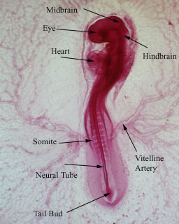
Figure 1. Stage 11 chick embryo, as determined by the Hamurger and Hamilton staging series (1951). Picture courtesy of Prof. Judy Cebra-Thomas.


Figure 2. Stage 14 chick embryo, as determined by the Hamburger and Hamilton staging series (1951). Picture courtesy of Prof. Judy Cebra-Thomas.


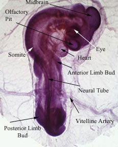
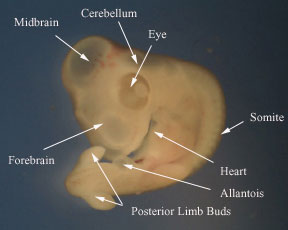
Figure 3. Stage 18 chick embryo, as determined by the Hamburger and Hamilton staging series (1951). Picture courtesy of Prof. Judy Cebra-Thomas.


Figure 4. Four-day old chick embryo. Picture courtesy of Anisha Chandra, Tuesday Developmental Biology Lab, Swarthmore College, PA, January 2004.


Pictures taken from Tuesday and Wednesday Developmental Biology Labs at Swarthmore College, PA, January 2004. In addition, some pictures were also courtesy of Prof. Judith CebraThomas from previous Developmental Biology classes.

Works Used in Labeling:

Cebra-Thomas, J. 2004. Observation of living and photography of chick embryos. Biology 24 Laboratory Handout. Swarthmore College, PA.

Gilbert, Scott. 2003. Developmental Biology, 7th ed. Sinauer Associates, Inc., MA, pp. 392, 398-399, 498-499, 526-528.