The amniote chicken egg looks unassuming, but is extremely cool. It contains
a huge single-celled yolk (the ovum) and enough water and nutrients to support
the developing chick until it hatches. Around 350 BC, Aristotle studied
the embryonic development of Gallus gallus, or the domestic chicken,
by cracking open an egg every day during the three-week incubation period.
He was the earliest known embryologist. Ever since then, the domestic chicken
has been a favorite model for studying vertebrate development. In 1951,
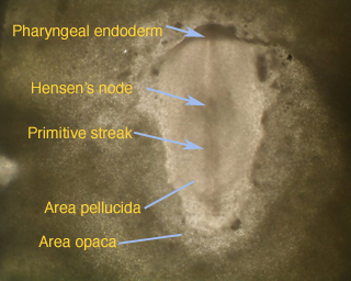
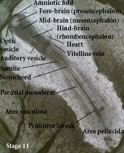
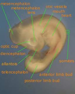
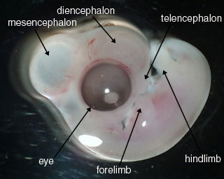
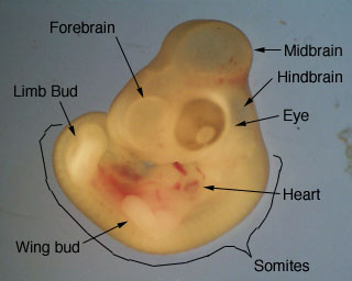
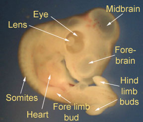
Hamburger and Hamilton took extremely detailed photos of chick development
and assigned stages to each visible step in the process. Below are pictures
of embryonic chicks at various HH stages of development (HH = Hamburger
and Hamilton), labeled by budding embryologists in the Swarthmore Developmental
Biology class.